39 human heart labelled diagram
› digestive-system-of-frogDigestive System of Frog | With Functions and Labelled Diagram Mar 06, 2022 · It is reddish-brown in color, the multilobed gland, which is situated close to the heart and lungs. The liver of frogs consists of 3 lobes –right, left, and median. The polygonal cells of the liver secrete a greenish alkaline fluid called bile. Human Heart Labeled Diagram The Human Heart Diagram Labeled - Human ... Jun 29, 2017 - Human Heart Labeled Diagram The Human Heart Diagram Labeled - Human Anatomy photo, Human Heart Labeled Diagram The Human Heart Diagram Labeled - Human Anatomy image, Human Heart Labeled Diagram The Human Heart Diagram Labeled - Human Anatomy gallery
Label the heart — Science Learning Hub In this interactive, you can label parts of the human heart. Drag and drop the text labels onto the boxes next to the diagram. Selecting or hovering over a box will highlight each area in the diagram. Right ventricle Right atrium Left atrium Pulmonary artery Left ventricle Pulmonary vein Semilunar valve Vena cava Aorta Download Exercise Tweet

Human heart labelled diagram
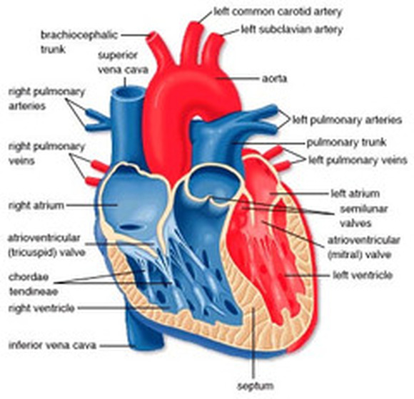
Human Heart Diagram - Human Body Pictures - Science for Kids Photo description: This is an excellent human heart diagram which uses different colors to show different parts and also labels a number of important heart component such as the aorta, pulmonary artery, pulmonary vein, left atrium, right atrium, left ventricle, right ventricle, inferior vena cava and superior vena cava among others. GCSE BIOLOGY HT 2022 ORGANISATION EXAM QA Q1. The diagram in Figure 1 shows a section through the human heart, seen from the front. Figure 1 (a) Draw a ring around the correct answer to complete each sentence. (i) The wall of the heart is made mostly of epithelial glandular muscular tissue. (1) (ii) The resting heart rate is controlled by the pacemaker. The pacemaker is located at position Human Heart - Anatomy, Functions and Facts about Heart The human heart is divided into four chambers, namely two ventricles and two atria. The ventricles are the chambers that pump blood and atrium are the chambers that receive the blood. Among which, the right atrium and ventricle make up the "right portion of the heart", and the left atrium and ventricle make up the "left portion of the heart." 5.
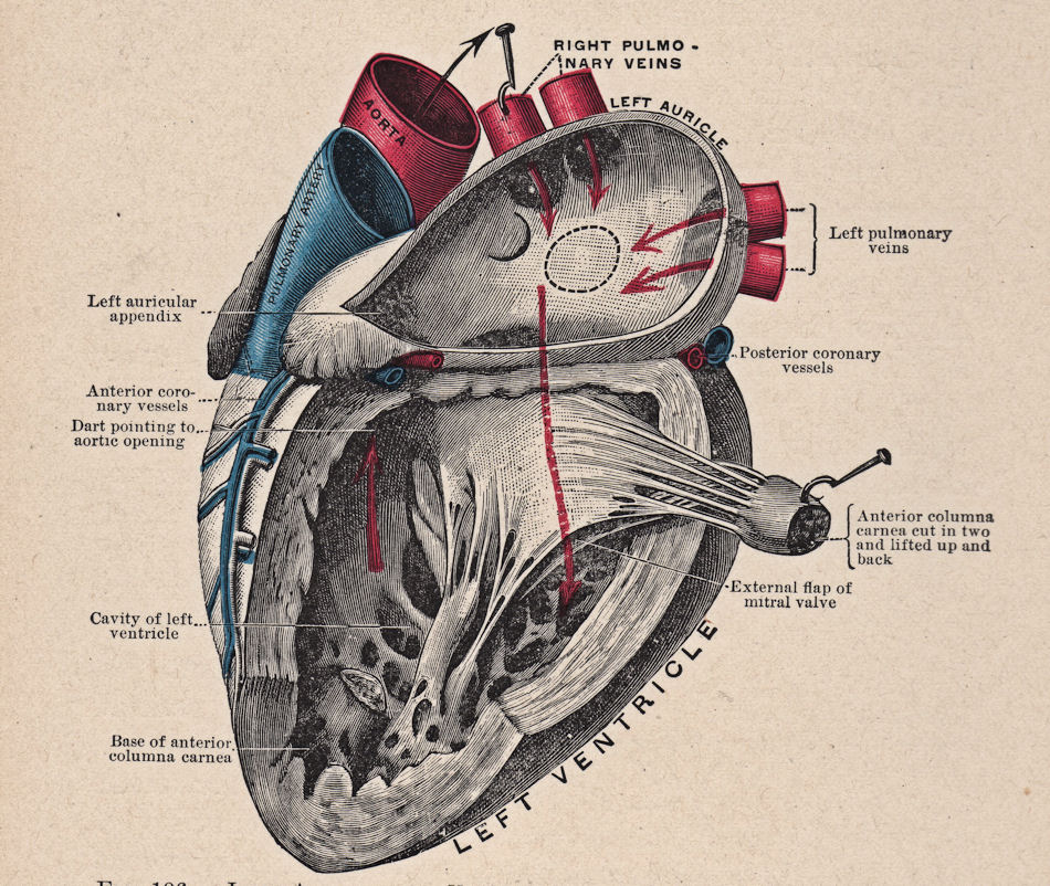
Human heart labelled diagram. commons.wikimedia.org › wiki › File:Diagram_of_theFile:Diagram of the human heart (cropped).svg - Wikimedia Apr 05, 2022 · English: Diagram of the human heart 1. Superior vena cava 2. 4. Mitral valve 5. Aortic valve 6. Left ventricle 7. Right ventricle 8. Left atrium 9. Right atrium 10. Aorta 11. Pulmonary v › photos › muscular-systemMuscular System Labeled Diagram Stock Photos, Pictures ... Human Heart labelled The heart is a hollow muscular organ that pumps blood throughout the blood vessels to various parts of the body by repeated, rhythmic contractions. muscular system labeled diagram stock pictures, royalty-free photos & images Labelled Heart Display Poster | Primary Resources | Twinkl A helpful labelled diagram of the human heart for display. This handy Labelled Heart Display Poster uses wonderful illustrations to show the main components of the heart. It's the perfect resource for a brilliant science display in your classroom, to give students a constant reference point when learning about the heart and its functions. Diagram of Human Heart and Blood Circulation in It Exterior of the Human Heart A heart diagram labeled will provide plenty of information about the structure of your heart, including the wall of your heart. The wall of the heart has three different layers, such as the Myocardium, the Epicardium, and the Endocardium. Here's more about these three layers. Epicardium
File:Diagram of the human heart (cropped).svg - Wikipedia File:Diagram of the human heart (cropped).svg. Size of this PNG preview of this SVG file: 611 × 600 pixels. Other resolutions: 244 × 240 pixels | 489 × 480 pixels | 782 × 768 pixels | 1,043 × 1,024 pixels | 2,086 × 2,048 pixels | 663 × 651 pixels. This is a file from the Wikimedia Commons. Information from its description page there is ... › human-body-organs-diagramHuman Body Organs Diagram Stock Photos and Images - Alamy Find the perfect human body organs diagram stock photo. Huge collection, amazing choice, 100+ million high quality, affordable RF and RM images. No need to register, buy now! human heart - Labelled diagram human heart - Labelled diagram Superior vena cava, inferior vena cava, Pulmonary vein, Pulmonary artery, Aorta, Tricuspid valve, Bicuspid valve, Septum, Right Atrium, Left Atrium, Right Ventricle, Left Ventricle. human heart Share by Mystudent78 Like Edit Content More Leaderboard Log in required Theme Log in required Options Switch template en.wikipedia.org › wiki › HeartHeart - Wikipedia The human heart is situated in the mediastinum, at the level of thoracic vertebrae T5-T8. A double-membraned sac called the pericardium surrounds the heart and attaches to the mediastinum. The back surface of the heart lies near the vertebral column, and the front surface sits behind the sternum and rib cartilages.
Human Heart (Anatomy): Diagram, Function, Chambers, Location in Body The heart is a muscular organ about the size of a fist, located just behind and slightly left of the breastbone. The heart pumps blood through the network of arteries and veins called the... Human Heart Diagram - Side View and Top View As shown in the human heart diagram above, the heart has several cavities (ventricles and atrium) and valves (pulmonary, aortic, mitral, tricuspid) which move blood in-and-out of the heart. It's pretty amazing to watch blood flow through the heart. To see how blood flows through the heart using an animation, please click here . Human Heart Diagram Illustrations, Royalty-Free Vector ... - iStock Browse 2,266 human heart diagram stock illustrations and vector graphics available royalty-free, or search for heart illustration or pulmonary artery to find more great stock images and vector art. heart anatomy. Part of the human heart. Human heart cross section, cardiovascular system diagram isolated on white. Heart Labeling Quiz: How Much You Know About Heart Labeling? Here is a Heart labeling quiz for you. The human heart is a vital organ for every human. The more healthy your heart is, the longer the chances you have of surviving, so you better take care of it. Take the following quiz to know how much you know about your heart. Questions and Answers. 1.
13+ Wall Of The Heart Diagram | Robhosking Diagram
Muscular System Labeled Diagram Pictures, Images and Stock … Human Heart labelled The heart is a hollow muscular organ that pumps blood throughout the blood vessels to various parts of the body by repeated, rhythmic contractions. muscular system labeled diagram stock pictures, royalty-free photos & images

Psychology | Mrs. Ayoub
drbiology001.files.wordpress.com › 2022 › 02GCSE BIOLOGY HT 2022 ORGANISATION EXAM QA Q1. - WordPress.com The diagram in Figure 1 shows a section through the human heart, seen from the front. Figure 1 (a) Draw a ring around the correct answer to complete each sentence. (i) The wall of the heart is made mostly of epithelial glandular muscular tissue. (1) (ii) The resting heart rate is controlled by the pacemaker. The pacemaker is located at position

a Lebelled Diagram of the Heart : Anatomy and physiology Heart - Cancer ...
Heart Diagram with Labels and Detailed Explanation - BYJUS The diagram of heart is beneficial for Class 10 and 12 and is frequently asked in the examinations. A detailed explanation of the heart along with a well-labelled diagram is given for reference. Well-Labelled Diagram of Heart. The heart is made up of four chambers: The upper two chambers of the heart are called auricles. The lower two chambers ...
Free Vintage Clip Art - Anatomy Heart - The Graphics Fairy
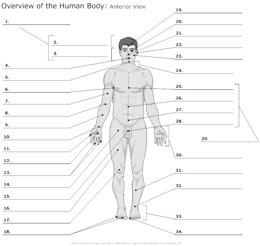
Organ Map | Diagram of Human Body Internal Organs Functions Featuring an accurate illustration and informative, clear labels, this organ map is a fantastic visual aid to support your teaching during science lessons all about the human body's internal organs.Once downloaded, you'll have an A4 diagram of a human body and internal organs, clearly labelled and perfect for individual use. Each internal organ is labelled and includes a …

Chordae tendineae - Wikipedia
Human Heart Diagram Labeled | Science Trends Human Heart Diagram Labeled Daniel Nelson 1, January 2019 | Last Updated: 3, March 2020 The human heart is an organ responsible for pumping blood through the body, moving the blood (which carries valuable oxygen) to all the tissues in the body. Without the heart, the tissues couldn't get the oxygen they need and would die.

Label Function Human Heart Diagram And Function - Aflam-Neeeak
Heart Anatomy: Labeled Diagram, Structures, Blood Flow ... - EZmed Image: Cardiac anatomy diagram showing the right and left side of the heart. The right side includes chambers 1 and 2. The left side includes chambers 3 and 4. Top vs Bottom of the Heart Next, we can divide the top 2 chambers of the heart from the bottom 2 chambers. The 2 chambers on top are known as the atria, and they include boxes 1 and 3.
Free Unlabelled Diagram Of The Heart, Download Free Unlabelled Diagram ...
Diagram of heart/How to draw human heart easily/human heart ... - YouTube Please watch: "cell structure and functions / animal cell vs plant cell / parts of cell / ch 8 science class 8 cbse" ...

Label parts of the heart interactive and downloadable worksheet. You ...
Describe the structure of the human heart with a neat labelled diagram. 1. Visceral Layer: It lines the outer surface of the heart. 2. Parietal Layer: It forms a sac which contains the fluid in the pericardial cavity around the outer region of the heart. (II) Heart Wall Structure: The wall of the heart consists of 3 layers, namely: -Epicardium - It is the outermost layer of the heart, composed of a thin-layered ...

Cross Sections Through the Thorax
Human Heart - Diagram and Anatomy of the Heart - Innerbody The heart is a muscular organ about the size of a closed fist that functions as the body's circulatory pump. It takes in deoxygenated blood through the veins and delivers it to the lungs for oxygenation before pumping it into the various arteries (which provide oxygen and nutrients to body tissues by transporting the blood throughout the body).
Heart Diagram - The Human Heart
A Diagram of the Heart and Its Functioning Explained in Detail The heart blood flow diagram (flowchart) given below will help you to understand the pathway of blood through the heart.Initial five points denotes impure or deoxygenated blood and the last five points denotes pure or oxygenated blood. 1.Different Parts of the Body ↓ 2.Major Veins ↓ 3.Right Atrium ↓ 4.Right Ventricle ↓ 5.Pulmonary Artery ↓ 6.Lungs

Circulatory System
13+ Heart Diagram Templates - Sample, Example, Format Download Now, instead of telling your students the heart ahs an artery in plain words, think of using a heart diagram template to make them understand what you are trying to tell them. ... All the minute parts inside the human heart have been clearly labelled. Free Download. Heart Structure And Its Functions Pdf Format.

Habits of the Heart: Lessons: Lung Diagram
byjus.com › biology › diagram-of-heartHeart Diagram with Labels and Detailed Explanation - BYJUS The diagram of heart is beneficial for Class 10 and 12 and is frequently asked in the examinations. A detailed explanation of the heart along with a well-labelled diagram is given for reference. Well-Labelled Diagram of Heart. The heart is made up of four chambers: The upper two chambers of the heart are called auricles. The lower two chambers ...

heart diagram unlabeled - Google Search | Heart diagram, Human heart ...
A Labeled Diagram of the Human Heart You Really Need to See The human heart, comprises four chambers: right atrium, left atrium, right ventricle and left ventricle. The two upper chambers are called the left and the right atria, and the two lower chambers are known as the left and the right ventricles. The two atria and ventricles are separated from each other by a muscle wall called 'septum'.
Post a Comment for "39 human heart labelled diagram"