41 photomicrograph of thick skin
Skin: The Histology Guide Dermis: Thick skin has a thinner dermis than thin skin, and does not contain hairs, sebaceous glands, or apocrine sweat glands. Thick skin is only found in areas where there is a lot of abrasion - fingertips, palms and the soles of your feet. show labels This is a picture of an H&E stained section of the epidermis of thin skin. microbewiki.kenyon.edu › index › Clostridium_perClostridium perfringens - microbewiki Apr 22, 2011 · It possesses the typical characteristics of Gram-positive bacteria, such as a protective thick cell wall, which is made up of peptidoglycan, surrounding an inner membrane. C. perfringens is an anaerobic bacterium, who acquires energy by performing anaerobic respiration using Nitrate as its electron acceptor. There is an increase in growth when ...
In the photomicrograph shown below which layer is only seen in thick ... Which statement best describes why the diagram represents thick skin? Choose the best answer. a) Thick skin lacks melanocytes. b) Thick skin contains five stratums. c) Thick skin epithelia is vascular. d) Thick skin epithelia is avascular. e) Thick skin contains stem cells. Answer: b lOMoARcPSD|9728372
Photomicrograph of thick skin
Solved The photomicrograph of thick skin | Chegg.com The photomicrograph of thick skin ; Question: The photomicrograph of thick skin . This problem has been solved! See the answer See the answer See the answer done loading. Show transcribed image text Expert Answer. Who are the experts? Experts are tested by Chegg as specialists in their subject area. We review their content and use your feedback ... photomicrograph of the epidermal layer in thick skin - Quizlet photomicrograph of skin. 5 terms. abba_dabba_17. skin and accessory structures diagram. 14 terms. shannon-ostenby PLUS. Epithelial tissue A. 12 terms. abba_dabba_17. Chapter 6 Flashcards | Quizlet In the photomicrograph shown below, which layer is only seen in thick skin? B. In the photomicrograph shown below, which layer do new cells arise? E. In the diagram of a hair root shown, which area is composed of dense connective tissue? F.
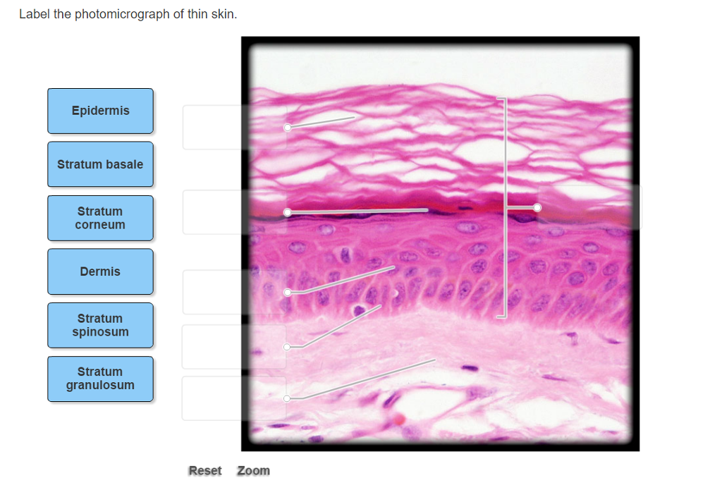
Photomicrograph of thick skin. 5.1 Layers of the Skin - Anatomy & Physiology Skin that has four layers of cells is referred to as "thin skin.". From deep to superficial, these layers are the stratum basale, stratum spinosum, stratum granulosum, and stratum corneum. Most of the skin can be classified as thin skin. "Thick skin" is found only on the palms of the hands and the soles of the feet. Solved Label the photomicrograph of thick skin | Chegg.com Label the photomicrograph of thick skin ; Question: Label the photomicrograph of thick skin . This problem has been solved! See the answer See the answer See the answer done loading. Show transcribed image text Expert Answer. Who are the experts? Experts are tested by Chegg as specialists in their subject area. We review their content and use ... photomicrograph of thick skin Diagram | Quizlet Start studying photomicrograph of thick skin. Learn vocabulary, terms, and more with flashcards, games, and other study tools. Anatomy and Physiology Homework Chapter 6 Flashcards - Quizlet The stratum granulosum consists of three to five layers of flat keratinocytes—more in thick skin than in thin skin. The stratum lucidum is a thin zone superficial to the stratum granulosum, seen only in thick skin. ... Label the photomicrograph of thick skin.-Stratum corneum-Stratum granulosum-Stratum spinosum-Stratum basale-Epidermis-Dermis ...
Label The Photomicrograph Of Thick Skin / Collagen Elastin Like ... Start studying photomicrograph of thick skin. "thick skin" is found only on the palms of the hands and the soles of the feet. Take several photomicrographs of thin skin at this magnification. A few layers of cells that are . Ready to take action to eliminate some wrinkles and defeat the signs of aging? staff-old.najah.edu › sites › defaultPhysiology and Anatomy of the Endocrine System Thick dry skin Puffy eyes Edema ... Photomicrograph of parathyroid gland tissue (1603). 49 . The Parathyroid Glands • They secrete parathyroid hormone 09 Histology of skin/How to Draw Thick Skin/Exams Preps Part B About Press Copyright Contact us Creators Advertise Developers Terms Privacy Policy & Safety How YouTube works Test new features Press Copyright Contact us Creators ... › pmc › articlesCT and MRI of adrenal gland pathologies - PMC Sep 13, 2018 · (G) Photomicrograph (original magnification, ×100; H-E stain) shows fibrous tissue, adipose tissue and muscle fibers are found in tumor tissues, and necrosis and calcification are noted. Hemorrhage Adrenal hemorrhage most commonly appears during the neonatal period and is rarely seen in adults ( 16 ), and can be divided into traumatic causes ...
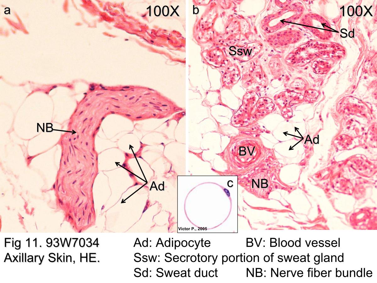
Solved Label the photomicrograph of thick skin. Epidermis | Chegg.com Expert Answer. Answer - There are two types of skin in human body : Thick skin Thin skin …. View the full answer. Transcribed image text: Label the photomicrograph of thick skin. Epidermis Stratum Basale Stratum lucidum Stratum cometim Stratum spinosu Stratus grinulosum Stratum corneum 10 Stratum lucidum Stratum granulosum Stratum spinosum ... Block1/Fig 11. Hypodermis of the thick skin. Fig 11. Hypodermis of the thick skin. The lower magnification photomicrograph shows part of the hypodermisof the thick skin. It contains abundant adipocytes. Theadipocyte (Ad) nucleus is compressed and displaced to oneside of the stored lipid droplets and the cytoplasm includingorganelles is reduced to a small rim (Fig 11c). Fig 11ashows several adipocytes and nerve fiber bundles (NB).Fig 11b ... en.wikipedia.org › wiki › Elastofibroma_dorsiElastofibroma dorsi - Wikipedia Elastofibroma dorsi is an ill-defined fibroelastic tumor-like condition made up of enlarged and irregular elastic fibers. The World Health Organization, 2020, has classified elastofibroma tumors as one specific type of the fibroblastic and myofibroblastic tumors. SKIN | The Big Picture: Histology | AccessBiomedical Science | McGraw ... Epidermis in thick skin, the type of skin found on the palms, flexor surfaces of the digits, and the soles of feet, is 400 to 600-μm thick. In comparison, epidermis in thin skin, which is found everywhere else on the body, is only 75 to 150-μm thick. Figure 11-1:
32 Label The Photomicrograph Of Thin Skin - Labels Design Ideas 2020
Block1/Fig 10. Dermis of thick skin - Kaohsiung Medical University Fig 10. Dermis of thick skin. This photomicrograph showsthe connective tissue of the skin, referred to as dermis,stained to show the nature and distribution of the elasticfibers (EF), which appear purple. The collagen fibers (CF)have been stained by eosin, and the two fiber types are easilydifferentiated. The elastic fibers of the dermis have a 3Dinterlacing configuration, thus the variety of ...
Biology Archive | January 15, 2017 | Chegg.com
› pmc › articlesMR imaging of ovarian masses: classification and differential ... Dec 16, 2015 · It is a benign germ cell tumour consisting of at least two of the three embryogenic germ cell layers, and usually contains ectodermal (skin, brain), mesodermal (fat, bone) and/or endodermal (thyroid tissue, gastrointestinal and bronchial epithelium) mature tissue . Simultaneous presence of these components leads to a complex and heterogeneous ...
Block1/Fig 11. Hypodermis of the thick skin.
Photomicrograph of Thick Skin Quiz - PurposeGames.com This is an online quiz called Photomicrograph of Thick Skin. There is a printable worksheet available for download here so you can take the quiz with pen and paper. Your Skills & Rank. Total Points. 0. Get started! Today's Rank--0. Today 's Points. One of us! Game Points. 6.

Ocular Pathology: Tissue Types-Epithelium, Blood Elements, Muscle etc.
Figure 7.1: Photomicrograph of Skin Diagram | Quizlet Start studying Figure 7.1: Photomicrograph of Skin. Learn vocabulary, terms, and more with flashcards, games, and other study tools.

35 Label The Photomicrograph Of Thin Skin. - Labels Information List
In the photomicrograph of a portion of thick skin - Course Hero Section Reference 1: Sec 5.1 Structure of the Skin. 33) In the photomicrograph of a portion of thick skin shown below, which layer is the stratum basale? a) Ab) B c) D d) Ee) F Answer: d. Difficulty: Medium Study Objective 1: SO 5.1 Describe the general structure of the skin.

Skin Showing Hair Follicles Sebaceous Glands Stock Photo 70287406 ...
Photomicrograph of Thick Skin - Printable - PurposeGames.com About this Worksheet. This is a free printable worksheet in PDF format and holds a printable version of the quiz Photomicrograph of Thick Skin.By printing out this quiz and taking it with pen and paper creates for a good variation to only playing it online.

Epidermal Layers. Mnemonic - Come, let's grow some berries ...
Label The Photomicrograph Of Thin Skin Quizlet - Skin Labeling Review ... Label the photomicrograph of thick skin. Learn vocabulary, terms, and more with flashcards, games, and other study tools. D) stratum corneum has fewer layers in. Label the photomicrograph of thin skin. C) contains more sweat glands than thin skin.

Chapter 12, Page 3 - HistologyOLM 4.0
› exotic-and-laboratoryParasitic Diseases of Fish - Exotic and Laboratory Animals ... Photomicrograph of gill tissue from a largemouth bass infected with Branchiomyces. Branching filaments of Branchiomyces can be easily seen in the gill tissue. The oval surrounded by a thick black ring is an air bubble. Unstained wet mount; original magnification, 100×.

Photomicrograph of Thick Skin
Low Magnification Micrograph Human Thin Skin Stock Photo ... - Shutterstock Download for free. Royalty-free stock photo ID: 1136062721. Low magnification micrograph of a human thin skin showing the epidermis and a very thick dermis, characteristic of this type of skin. Hematoxylin-eosin.
Post a Comment for "41 photomicrograph of thick skin"