43 cell diagram to label
Label the cell diagram - Teaching resources - Wordwall Copy of Plant Cell Diagram Labeling Labelled diagram. by Swilliams9. 5.6 Label the sentence Labelled diagram. by Christianjolene. Label the 3D Shapes Labelled diagram. by Erinbecerra. G1 shapes 3D Shapes. Label the Electromagnetic Spectrum Labelled diagram. by Elizabetheck. G6 G7 G8 Science. 03 Label the Cell Diagram | Quizlet Biology Cell Biology 03 Label the Cell STUDY Learn Flashcards Write Spell Test PLAY Match Gravity Created by muskopf1TEACHER Terms in this set (14) Nucleus Control center of the cell Nucleolus Ribosome synthesis Rough Endoplasmic Reticulum Protein transport Smooth Endoplasmic Reticulum Lipid synthesis Mitochondrion Cellular Respiratoin
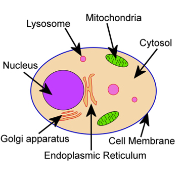
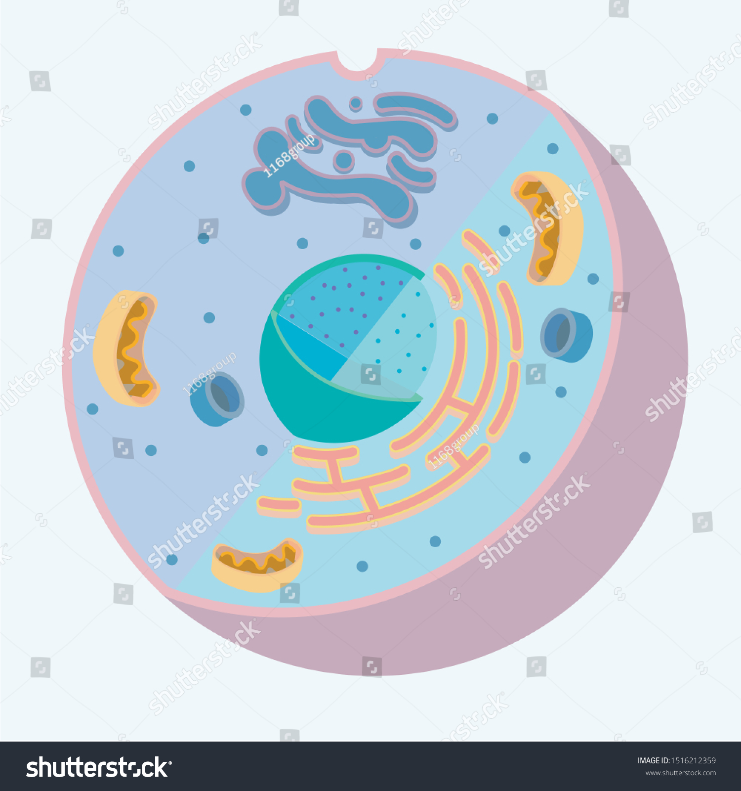
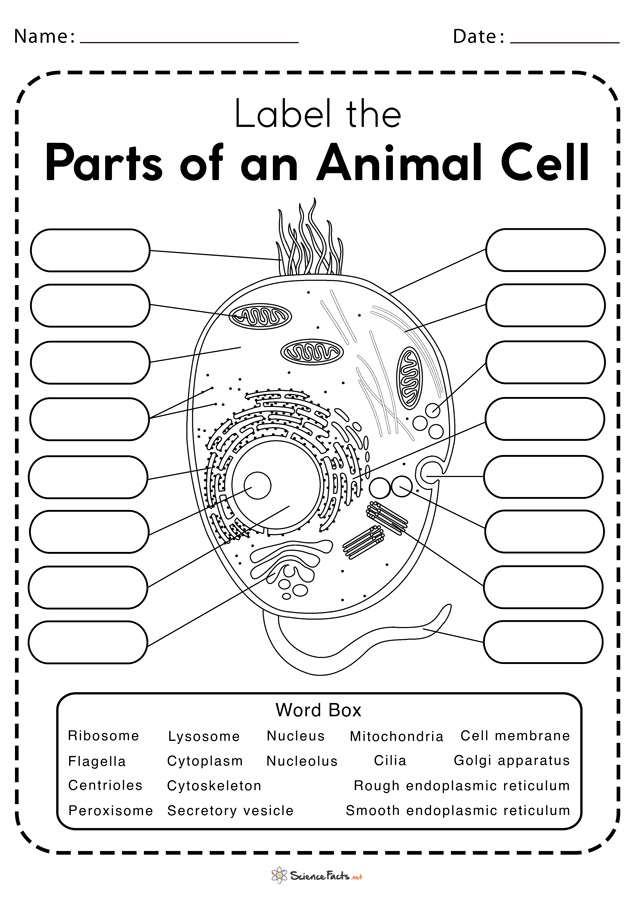
Animal Cell Diagram with Label and Explanation: Cell ... - Collegedunia Diagram of Animal Cell Below is the diagram of the animal cell which shows the organelles present in it. The cell is covered with cytoplasm which consists of cell organelles in it. The nucleus is covered with a rough Endoplasmic Reticulum and other organelles each designed for a specific purpose.

Cell diagram to label
Cell Diagrams with Labelling Activity | Learnful The cell structure illustrations for these diagrams were generated in BioRender. Both diagrams feature a drag-and-drop labelling activity created with H5P here ... Learn the parts of a cell with diagrams and cell quizzes Cell diagram unlabeled It's time to label the cell yourself! As you fill in the cell structure worksheet, remember the functions of each part of the cell that you learned in the video. Doing this will help you to remember where each part is located. Click the links below to download the labeled and unlabeled eukaryotic cell diagrams. cell diagrams to label cell diagrams to label IB Biology Notes - 2.3 Eukaryotic cells. 8 Images about IB Biology Notes - 2.3 Eukaryotic cells : Plant and Animal Cell Labeling (Color), Chromosome Diagrams and also Chromosome Diagrams. IB Biology Notes - 2.3 Eukaryotic Cells ibguides.com eukaryotic electron cell liver micrographs micrograph ultrastructure
Cell diagram to label. Animal Cells: Labelled Diagram, Definitions, and Structure - Research Tweet Animal Cells: Labelled Diagram, Definitions, and Structure What is a Cell? In biology, cell is the smallest unit that can live on its own and that makes up all living organisms and the tissues of the body. A cell has three main parts: the cell membrane, the nucleus, and the cytoplasm. What is Animal Cell? Plant and Animal Cell: Labeled Diagram, Structure, Function - Embibe Plant Cell: Plant cells are eukaryotic cells with a true nucleus along with specialized structures called organelles that carry out certain specific functions. Animal Cell: An animal cell is a type of eukaryotic cell that lacks a cell wall and has a true, membrane-bound nucleus along with other cellular organelles. Diagram of Plant and Animal Cell blank cell diagram to label cell animal labeled cells diagram structure typical celula para imagen 3d biology labels figure membrane parts label organelles function discovery. Label The Cell Diagram . labeling labeled organelles exploringnature diagrams 2nd. 15 Best Images Of Human Anatomy Physiology Worksheets - Unlabeled Cell Labeling Quiz - PurposeGames.com This is an online quiz called Cell Labeling. There is a printable worksheet available for download here so you can take the quiz with pen and paper. Your Skills & Rank. Total Points. 0. Get started! Today's Rank--0. Today 's Points. One of us! Game Points. 10. You need to get 100% to score the 10 points available.
Get Blank Animal Cell Diagram To Label Pdf 2020-2022 - US Legal Forms Follow these simple steps to get Blank Animal Cell Diagram To Label Pdf ready for submitting: Choose the sample you require in the collection of templates. Open the document in the online editor. Read through the guidelines to find out which info you will need to give. Select the fillable fields and put the necessary info. Cell: Structure and Functions (With Diagram) - Biology Discussion Eukaryotic Cells: 1. Eukaryotes are sophisticated cells with a well defined nucleus and cell organelles. 2. The cells are comparatively larger in size (10-100 μm). 3. Unicellular to multicellular in nature and evolved ~1 billion years ago. 4. The cell membrane is semipermeable and flexible. 5. These cells reproduce both asexually and sexually. Red Blood Cell Diagram Labeled stock illustrations Red Blood Cell Diagram Labeled Illustrations, Royalty-Free Vector Graphics & Clip Art - iStock Choose from Red Blood Cell Diagram Labeled stock illustrations from iStock. Find high-quality royalty-free vector images that you won't find anywhere else. Video Trending searches Fathers day Summer background Cellulite Monkeypox Plants Coffee Converting Diagrams - The Biology Corner This will add text boxes that can be filled in. You can also use drop-down fields if you want to provide students with choices in how to label the diagram. Students will need to download the pdf in order to fill in the fields. Open Google Draw and import the diagram. Then use "insert" to create text boxes where students can fill in the labels.
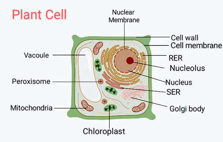
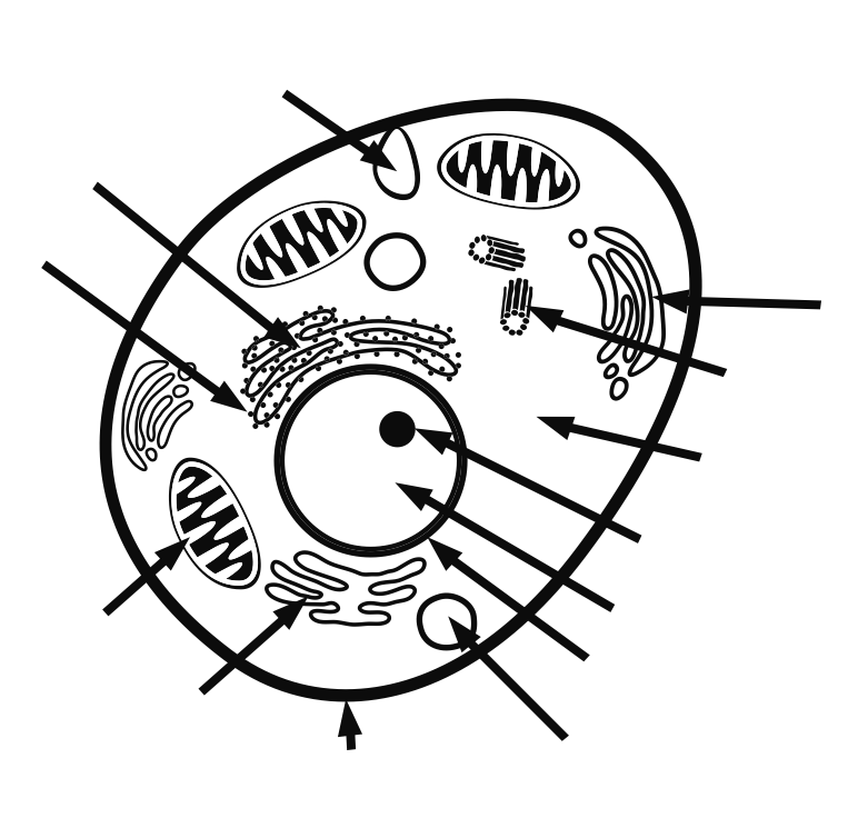
PDF Human Cell Diagram, Parts, Pictures, Structure and Functions Diagram of the human cell illustrating the different parts of the cell. Cell Membrane The cell membraneis the outer coating of the cell and contains the cytoplasm, substances within it and the organelle. It is a double-layered membrane composed of proteins and lipids. Cell Organelles- Definition, Structure, Functions, Diagram - Microbe Notes In a plant cell, the cell wall is made up of cellulose, hemicellulose, and proteins while in a fungal cell, it is composed of chitin. A cell wall is multilayered with a middle lamina, a primary cell wall, and a secondary cell wall. The middle lamina contains polysaccharides that provide adhesion and allow binding of the cells to one another. Labeled Plant Cell With Diagrams | Science Trends To start with, the entire cell is enveloped by a rigid structure called the cell wal l. The cell wall's function is to give protection to the cell and to support it. The cell wall must be both permeable yet rigid. It must be permeable so that materials can move in and out of the cell, but rigid enough that it supports and protects the cell. cell diagram labeling worksheet Skeleton bones worksheet human anatomy worksheets printable blank diagram labeled bone skeletal body system labeling appendicular unlabeled skull muscle physiology. 11 best images of blank anatomy worksheets cell diagram labeling worksheet. 12 Best Images of Compare And Contrast Worksheet Grade 1 - Parts of a. 9 Images about 12 Best Images of ...
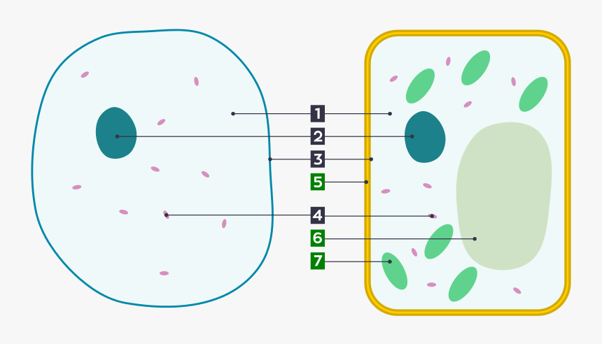
Plant and Animal Cells activity
Structure of Cell: Definition, Types, Diagram, Functions - Embibe Cell Definition Cells are the fundamental structural and functional unit of all living beings including plants, animals and microorganisms. All living organisms in this universe are made up of cells. We cannot see cells with naked eyes as they are only \ (10\) microns in size whereas human eyes cannot see objects less than \ (100\) microns.
Label animal cell - Teaching resources
Label that Diagram - Cells - Apps on Google Play There are 5 cells presented: Animal Cell, Plant Cell, Amoeba, Paramecium, and Euglena. The player can study the labeled diagrams or play the game of labeling the diagrams. When the game is played, the labels appear in a random order one at a time and the player must tap on the correct dot on the diagram.
Draw a diagram of a plant cell and label at least eight class ...
Interactive Cell Model - CELLS alive Cell Wall. Chloroplast. Smooth Endoplasmic Reticulum. Rough Endoplasmic Reticulum. Ribosomes. Cytoskeleton. RETURN to CELL DIAGRAM ...
Animal and Plant Cell Labeling
Label Cell Parts | Plant & Animal Cell Activity Students will create a cell diagram labeled with the different organelles of plant and animal cells. The cell diagrams are easily colorable, allowing students ...

Parts of plant cell worksheet
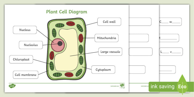
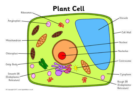
Plant Cells: Labelled Diagram, Definitions, and Structure - Research Tweet Plastids and Chloroplasts. Plants make their own food through photosynthesis. Plant cells have plastids, which animal cells don't. Plastids are organelles used to make and store needed compounds. Chloroplasts are the most important of plastids. They convert light energy from the sun into sugar and oxygen. The most exposed parts of the plants ...
Cell Diagrams with Labelling Activity | Learnful
Animal Cell Labelling Activity | Primary Resources - Twinkl A colorful resource which covers the parts of an animal cell including the nucleus, cell wall, cytoplasm, and mitochondria. Lower, middle and higher ability ...
Animal & Plant Cell worksheets: Colour & label | Teaching ...
A Labeled Diagram of the Animal Cell and its Organelles A Labeled Diagram of the Animal Cell and its Organelles There are two types of cells - Prokaryotic and Eucaryotic. Eukaryotic cells are larger, more complex, and have evolved more recently than prokaryotes. Where, prokaryotes are just bacteria and archaea, eukaryotes are literally everything else.

Animal and Plant Cells Worksheet New Gcse Cell Biology by ...
Free Cell Diagram Software with Free Templates - EdrawMax - Edrawsoft How to Make a Cell Diagram? Step 1: Initial working Before making a cell diagram on EdrawMax, first gather all the necessary supporting facts to draw the diagram. Draw all the cell components roughly into the shape of a cell. The cell wall, cell membrane, cytoplasm, nucleus, and cell organelles are components. Step 2: Template selection

Plant and Animal Cell Worksheets
Cell Worksheets | Plant and Animal Cells - Math Worksheets 4 Kids Plant Cell Diagram | Animal Cell Diagram. Featured in this printable worksheet are the diagrams of the plant and animal cells with parts labeled vividly. This enhanced visual instructional tool assists in grasping and retaining the names of the cell parts like mitochondrion, vacuole, nucleus and more with ease.
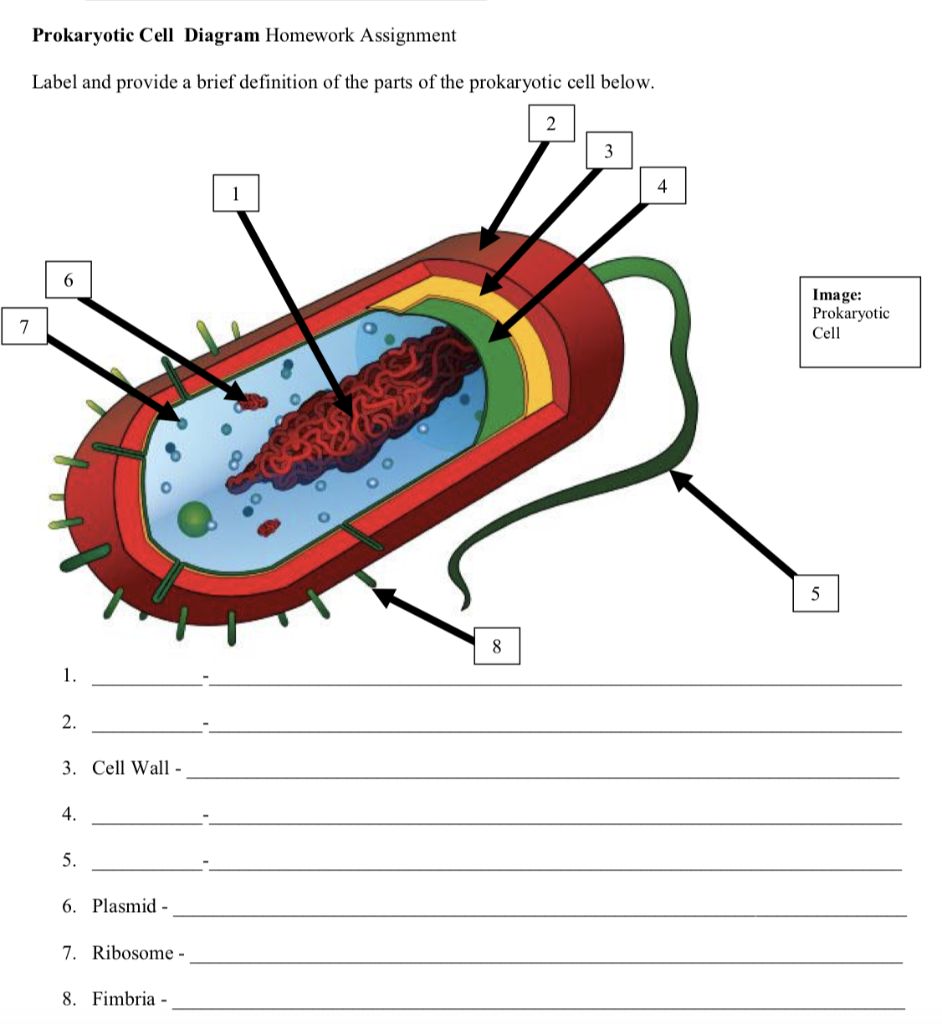
Solved Prokaryotic Cell Diagram Homework Assignment Label ...
Label the cell - Teaching resources - Wordwall Label Plant and Animal Cell Labelled diagram by Eawilson the cell Match up by Elenagp9149 5.6 Label the sentence Labelled diagram by Christianjolene Label the Electromagnetic Spectrum Labelled diagram by Elizabetheck G6 G7 G8 Science 5.7 Label the sentence Labelled diagram by Christianjolene The cell Anagram by Thepowerhouse G7 G8 Science
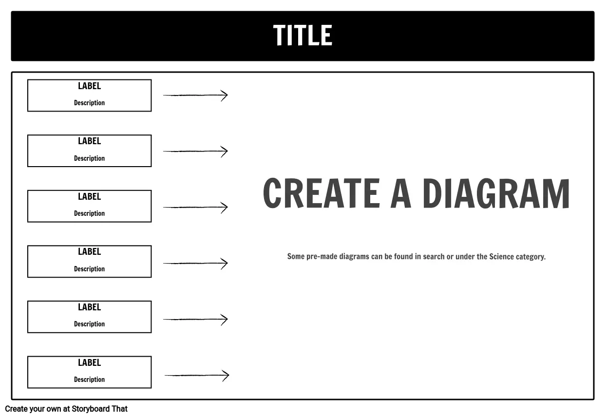
Label Cell Parts | Plant & Animal Cell Activity | StoryboardThat
Label that Diagram - Cells - Apps on Google Play 12 Oct 2019 — This is the 64-bit edition of the latest version of this app. Enjoy. This app provides the user the opportunity to study cell structure as ...
Animal Cell Without Labels - ClipArt Best
Cell Cycle Diagram Labeled | EdrawMax The following labeled diagram illustrates the cell cycle. As you can see in the labeled diagram here, the cell cycle is an ordered series of events involving cell growth and cell division that produces two new daughter cells. Cells on the path to cell division proceed through a series of precisely timed and carefully regulated stages of growth ...

CELLS Blank Plant & Animal Cell Diagrams: Note Taking ...
A Well-labelled Diagram Of Animal Cell With Explanation - BYJUS The animal cell diagram is widely asked in Class 10 and 12 examinations and is beneficial to understand the structure and functions of an animal. A brief explanation of the different parts of an animal cell along with a well-labelled diagram is mentioned below for reference. Also Read Different between Plant Cell and Animal Cell
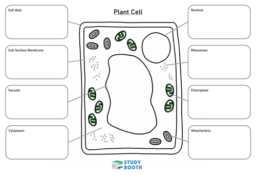
Labelled Plant Cell Diagram - Handy Worksheets for Children
Cell Diagram | Free Cell Diagram Templates - Edrawsoft A free customizable cells diagram template is provided to download and print. Quickly get a head-start when creating your own cell diagram. Here is a simple cell diagram example created by Science Diagram Maker Download Template: Get EdrawMax Now! Free Download Popular Latest Flowchart Process Flowchart Workflow BPMN Cross-Functional Flowchart

Animal Cell- Definition, Structure, Parts, Functions, Labeled ...
cell diagrams to label cell diagrams to label IB Biology Notes - 2.3 Eukaryotic cells. 8 Images about IB Biology Notes - 2.3 Eukaryotic cells : Plant and Animal Cell Labeling (Color), Chromosome Diagrams and also Chromosome Diagrams. IB Biology Notes - 2.3 Eukaryotic Cells ibguides.com eukaryotic electron cell liver micrographs micrograph ultrastructure

Label that Diagram - Cells - YouTube
Learn the parts of a cell with diagrams and cell quizzes Cell diagram unlabeled It's time to label the cell yourself! As you fill in the cell structure worksheet, remember the functions of each part of the cell that you learned in the video. Doing this will help you to remember where each part is located. Click the links below to download the labeled and unlabeled eukaryotic cell diagrams.
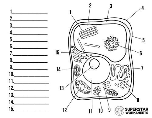
Plant Cell Worksheets - Superstar Worksheets
Cell Diagrams with Labelling Activity | Learnful The cell structure illustrations for these diagrams were generated in BioRender. Both diagrams feature a drag-and-drop labelling activity created with H5P here ...

parts of a cell label - Clip Art Library

Label the cell structure. | Study.com
Label the Plant Cell Worksheets (SB11867) - SparkleBox

Draw an outline diagram of an animal cell. Label its different parts. | 6 | THE CELL | BIOLOGY...

Converting Diagrams

Draw a diagram of typical cell and label the following parts ...

Animal Cell Diagram To Label - Biology Forums Gallery
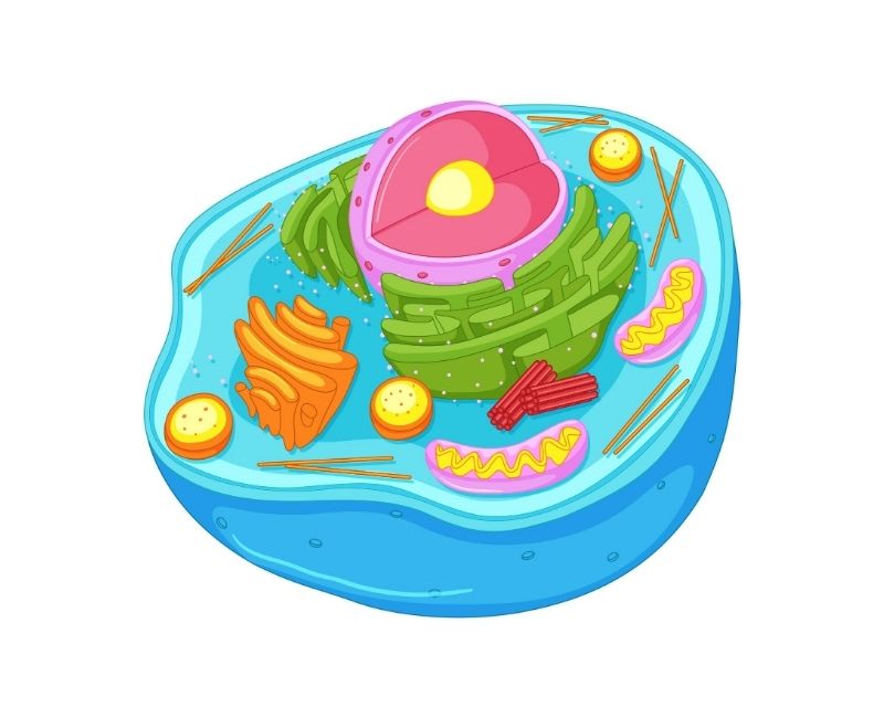
3d model of animal cell | Animal cell diagram to label worksheet
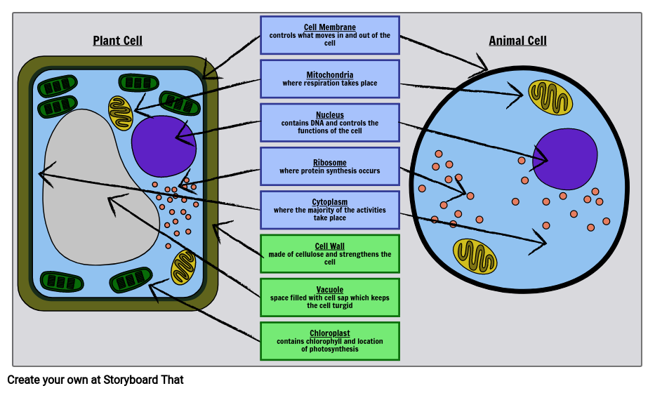
Label Cell Parts | Plant & Animal Cell Activity | StoryboardThat
Label animal cell - Teaching resources
Vektor Stok Diagram Animal Cell Organelle Without Labels ...

Plant Cell and Animal Cell Diagram Quiz
Basic Animal And Plant Cells Clipart , Png Download - Animal ...
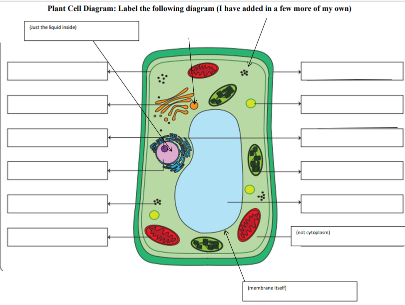
Solved Plant Cell Diagram: Label the following diagram (I ...
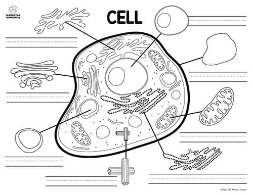
Animal Cell Worksheet - Superstar Worksheets

Label the Plant Cell Worksheet
Animal Cell - Free printable to label + Color -kidCourses.com

The Animal and Plant Cells Colour and Label Diagram ...

Label the following diagram of animal cell.Answer it ...
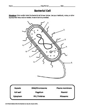
Bacterial Cell Labeling Diagram

Human Cell Coloring Page
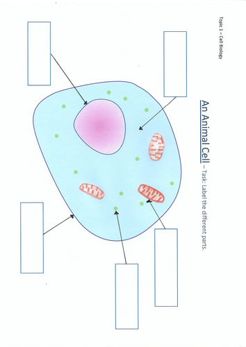
An Animal Cell diagram to label | Teaching Resources
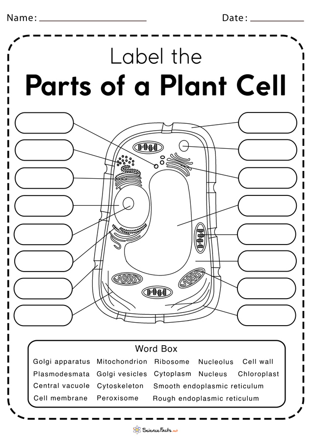
Plant Cell Worksheets - Free Printable
Label animal cell - Teaching resources
Animal Cell Worksheets - Free Printable

Label the Cell Diagram | Quizlet
Post a Comment for "43 cell diagram to label"